

Endodontics
Endodontic microsurgery
A targeted procedure at the tip of the root, when a conventional treatment isn't enough.
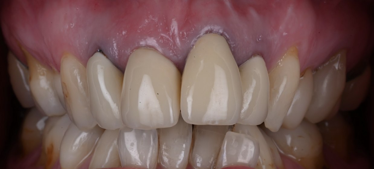
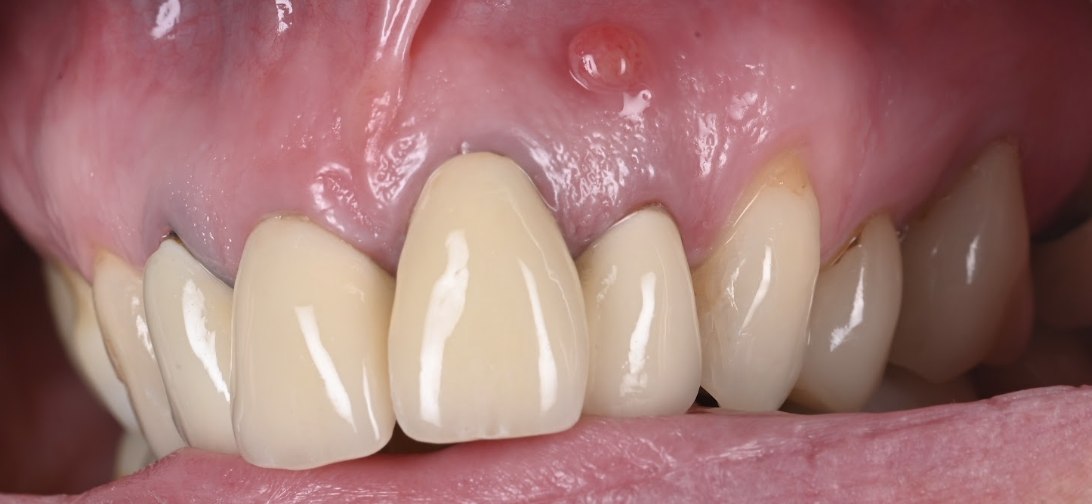
Endodontic microsurgery, or apicoectomy, is a procedure performed when a conventional endodontic (re)treatment is not possible or not advisable.
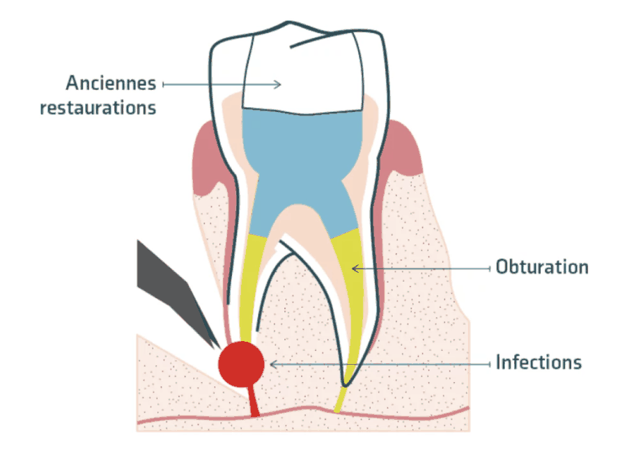
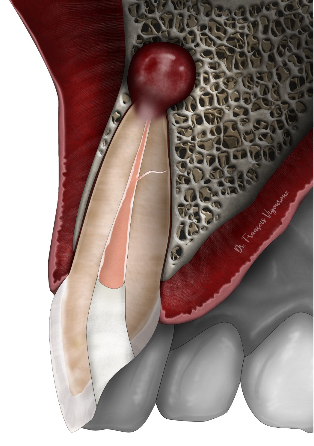
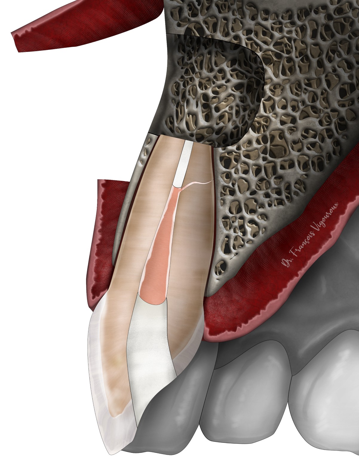
It involves accessing the tip of the root (apex) directly through minor surgery. The infected part of the bone (cyst or granuloma) and of the root is removed. The root is then cleaned and sealed from the opposite direction (retrograde approach) with a biocompatible material to prevent reinfection.
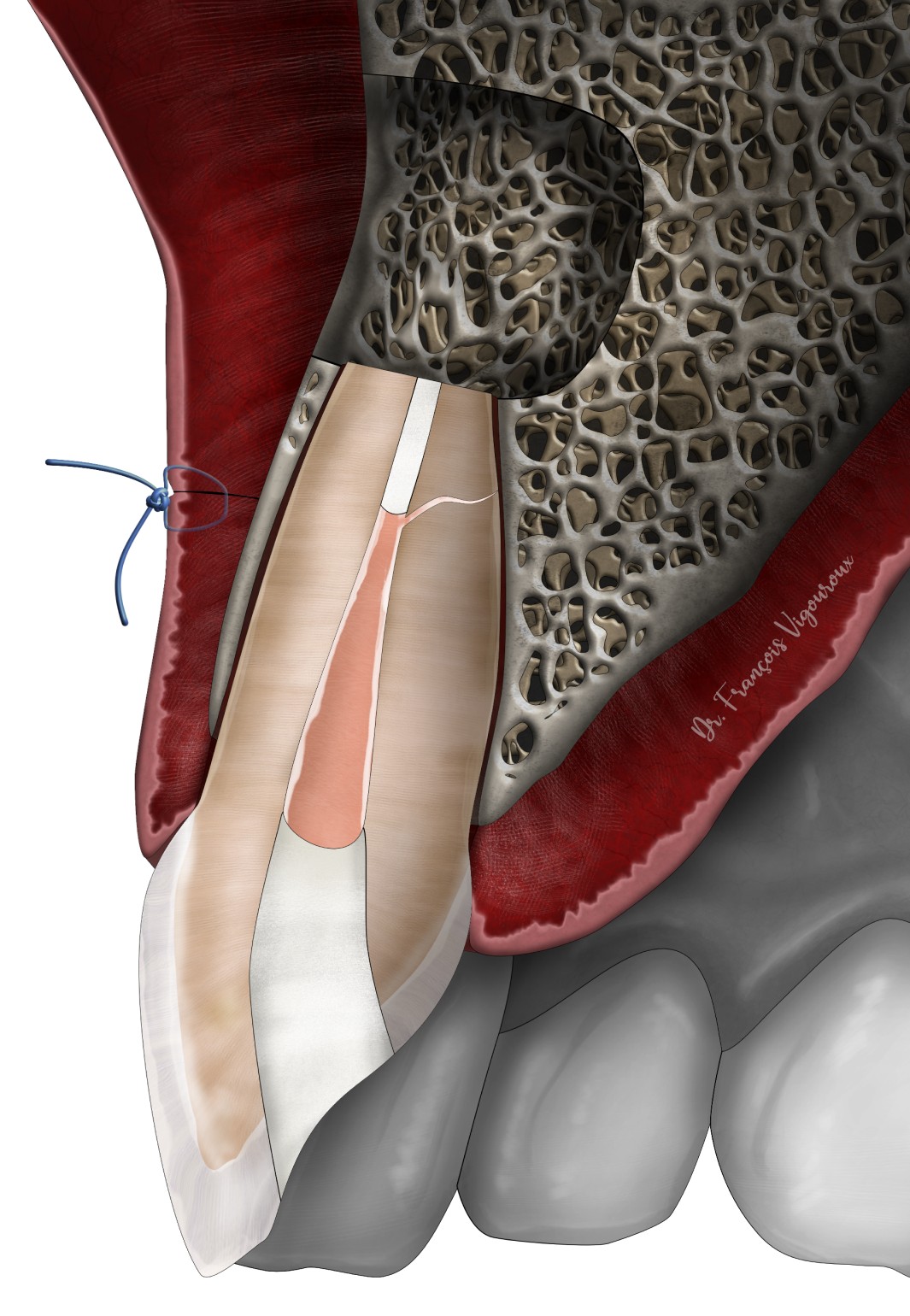
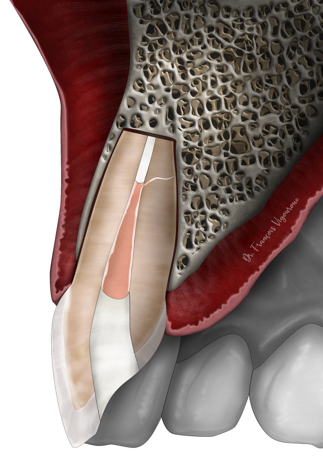
It is performed with the aid of optical magnification.
A question about this treatment?
Book an appointment, and we will answer all your questions during the consultation.